| News |
 |
|
|
|
|
|
|
|
|
| What Is The Aortic Valve? |
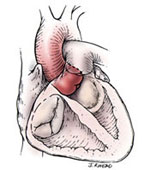
The location of the aortic valve is shown in the diagram to the left. The heart has two sets of pumping chambers: the right-sided chambers pump blood to the lungs, and the left side pumps blood to the rest of the body. The left side, therefore, has a harder job than the right side, and the left side does most of the work. The main pumping chambers of the heart are called the ventricles.
Because the ventricle is a pump, it must have both an inflow valve and an outflow valve. The aortic valve is on the left side of the heart and is the outflow valve. The aortic valve opens to allow blood to leave the left ventricle (the main pumping chamber of the heart) and closes to prevent blood from leaking backwards into the ventricle from the rest of the body. |
|
|
|
| What Causes An Aortic Valve To Malfunction? |
The aortic valve to malfunction for several reasons. For example, the aortic valve may be abnormal from birth (congenital aortic valve disease), or it could become diseased with age (acquired aortic valve disease).
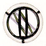
The most common congenital abnormality is a bicuspid aortic valve. As shown below, the aortic valve normally has three leaflets, but a bicuspid aortic valve has only two. It may, therefore, not open or close completely.
A bicuspid aortic valve is a common abnormality and occurs in 1-2% of people. This is the second most common cause of aortic valve disease requiring surgery. Such valves may function normally for years before becoming stenotic, regurgitant, or both. People with a bicuspid aortic valve require antibiotic prophylaxis before dental procedures but generally no other special precautions are required other than regular follow up with a qualified cardiologist.
|
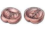 |
| Tricuspid (left) and Bicuspid (right) Aortic Valves |
|
|
The most common cause of aortic valve disease requiring surgery is called "senile aortic calcification," meaning that the valve has worn out with age. When a valve becomes worn, the body deposits calcium on it for reasons that are unknown. The calcium restricts or limits the motion of the valve leaflets. This may prevent the valve from opening (causing stenosis) or closing (causing leakage or regurgitation). Less common causes of aortic valve disease include diseases of the aorta, the main blood vessel coming out of the heart and carrying blood to the rest of the body, including ascending aortic aneurysms, aortic dissection, and Marfan's syndrome. |
|
Are There Any Warning Signs For A Failing Aortic Valve? |
A failing aortic valve may cause a variety of symptoms including shortness of breath, chest pain (angina pectoris), and dizziness or loss of consciousness (passing out).
A narrow valve makes the heart work harder just to pump the blood through the valve to the body. A leaky valve lets blood back into the heart after it has been pumped out. The heart must therefore pump more blood forward to make up for the blood that is leaking backwards. Either way the extra work may cause symptoms of heart failure, such as shortness of breath. Early on the shortness of breath may be noticeable only with exercise. Later, with the progression of valve disease, a patient could experience shortness of breath with even light activity or at rest. Some patients will be unable to sleep flat in bed or may awaken from sleep short of breath. Another sign of heart failure that may occasionally occur is swelling of your feet, particularly prominent later in the afternoon or evening although other conditions, such as varicose veins, can also cause this to occur.
The extra work the heart has to perform may also cause chest pain or angina pectoris similar to the symptoms of a heart attack. It may be difficult to tell the difference between heart valve disease and narrowing of the blood vessels to the heart itself (coronary arteries).
Aortic valve disease may also cause dizziness, light headedness or even fainting spells.
|
|
| How Does Someone Know If They Should Have Surgery To Repair An Aortic Valve? |
The decision to proceed with surgery should be made with your medical care team which usually consists of a thoracic or cardiothoracic surgeon and a cardiologist. Your medical team will likely base their recommendations on your symptoms and on the results of several tests including an echocardiogram and sometimes cardiac catheterization. An echocardiogram may show enlargement of the heart, and can help to measure the degree of stenosis or regurgitation. A cardiac catheterization provides similar information, but can also identify any narrowings of the coronary arteries. |
|
|
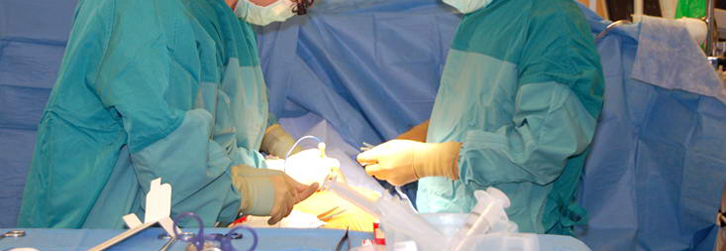
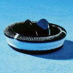
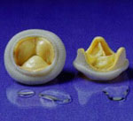
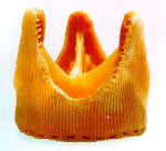
| What Options Exist For The Replacement Of Artificial Valves? |
Unlike the mitral valve which can often be repaired, the aortic valve usually requires replacement. Once the decision is made to proceed with surgery, choices regarding the type of artificial valve (prosthesis) used should be considered. In broad terms there are two types of artificial valves or prostheses: mechanical valves and biological valves. Examples of the valves that your cardiothoracic surgeon might use are pictured below. |
|
|
| Are There Differences Between Mechanical And Biological Heart Valves? |
A number of excellent mechanical replacement valves or prostheses are available today. Most surgeons have a particular preference for one valve over another related to technical factors (how they are sewn into place), however from the patient's point of view there is little if any difference between valves. The principle advantage of mechanical valves is their excellent durability. From a practical standpoint, they do not wear out. The principle disadvantage is that there is a tendency for blood to clot on all mechanical valves. Therefore patients with artificial valves must take anticoagulants or "blood thinners" for the rest of their life. There is also a small but definite risk of blood clots causing stroke. pictured below. |
|
There are a variety of natural or biological valves that can be used to replace an abnormal valve. They all share a reduced risk of blood clots forming but all are less durable than mechanical valves. Given enough time, they will probably all wear out. The options in this category include "xenograft" valves made from animal tissues (most often pig aortic valves), "homograft" or "allograft" valves retrieved from human cadavers, and "pulmonary autograft" valves moved from the patient's pulmonary artery on the right side of the heart to the aortic position on the left.
The decision on the type of valve used should be made in conjunction with your cardiothoracic surgeon and your cardiologist. Ultimately the choice will depend on a patient's preferences, lifestyle, and individual risks as determined by age and other medical conditions. |
|
| Why Is Surgery Necessary? |
The aortic valve is the outflow valve of the left side of the heart, meaning that it opens during systole (when the ventricle contracts or squeezes blood out into the aorta and the rest of the body). When the aortic valve is too narrow or stenotic, the ventricle has to work harder to pump the blood out to the body. This extra work consumes significant energy and ultimately requires extra blood flow to the heart itself. If there is not enough blood flow, the heart becomes ischemic causing anginal chest pain. Aortic stenosis is often progressive, growing worse with time. As the valve gets tighter, the heart has to continue to work harder and harder to keep pumping blood out of the heart. At some point the heart can no longer compensate, resulting in episodes of low blood pressure or hypotension or even syncope. As the heart fails to compensate, fluid will build up in the lungs creating congestion.
When the aortic valve leaks, the heart has to work harder and similar problem occur. The ventricle must pump more blood with each contraction to produce the same forward output, creating a condition called volume overload. The heart can compensate for this volume overload for many months or years provided the leakage develops slowly. Eventually, the heart begins to fail producing shortness of breath and fatigue.
|
|
| What Are The Risks Of Surgery? |
Individual risks of surgery can be best estimated by your cardiothoracic surgeon and cardiologist. Risks generally depend on age, general health, specific medical conditions, and heart function. |
|
| What Will My Condition Belike After Aortic Valve Replacement? |
After successful aortic valve replacement, patients can expect to return to their preoperative condition or better. Anticoagulation ("blood thinners") with a drug like Coumadin may be prescribed for 6 weeks to 3 months after surgery for those with biological valves, and for life for those with mechanical valves. Once the wounds have healed, most patients should experience few if any restrictions to activity. A patient will require preventative or prophylactic antibiotics whenever having dental work, and should always tell a doctor about their valve surgery before any surgical procedure |
|
|
|
|
|
|